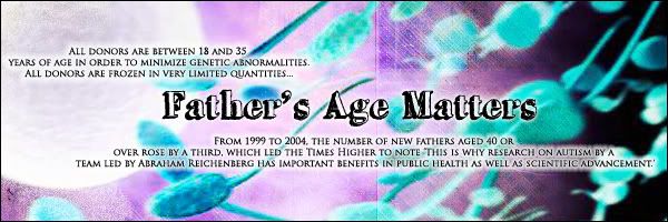
AUTISM PREVENTION FATHER BABIES 24-34 PATERNAL AGE IS KEY IN NON-FAMILIAL AUTISMVaccines

"It is very possible that PATERNAL AGE is the major predictor of(non-familial) autism." Harry Fisch, M.D., author "The Male Biological Clock". Sperm DNA mutates and autism, schizophrenia bipolar etc. results. What is the connection with autoimmune disorders? Having Type 1 diabetes, SLE,etc. in the family, also if mother had older father. NW Cryobank will not accept a sperm donor past 35th BD to minimize genetic abnormalities.VACCINATIONS also cause autism.

Friday, February 29, 2008

Below is a verbatim copy of the US Government concession filed last November in a vaccine-autism case in the Court of Federal Claims, with the names of the family redacted.t:http://www.huffingtonpost.com/david-kirby/the-vaccineautism-court-_b_88558.h


Every American should read this document, and interpret for themselves what they think their government is trying to say about the relationship, if any, between immunizations and a diagnosis of autism spectrum disorder.

If you feel this document suggests that some kind of link may be possible, you might consider forwarding it to your elected representatives for further investigation.

But, of course, if you feel that this document in no way implicates vaccines, then let’s just keep going about our business as usual and not pay any attention to all those sick kids behind the curtain.

IN THE UNITED STATES COURT OF FEDERAL CLAIMS
OFFICE OF SPECIAL MASTERS
CHILD, a minor,

by her Parents and Natural Guardians,

Petitioners,

v.

SECRETARY OF HEALTH AND HUMAN SERVICES,

Respondent.

RESPONDENT’S RULE 4(c) REPORT

In accordance with RCFC, Appendix B, Vaccine Rule 4(c), the Secretary of Health and Human Services submits the following response to the petition for compensation filed in this case.

FACTS

CHILD (”CHILD”) was born on December –, 1998, and weighed eight pounds, ten ounces. Petitioners’ Exhibit (”Pet. Ex.”) 54 at 13. The pregnancy was complicated by gestational diabetes. Id. at 13. CHILD received her first Hepatitis B immunization on December 27, 1998. Pet. Ex. 31 at 2.

From January 26, 1999 through June 28, 1999, CHILD visited the Pediatric Center, in Catonsville, Maryland, for well-child examinations and minor complaints, including fever and eczema. Pet. Ex. 31 at 5-10, 19. During this time period, she received the following pediatric vaccinations, without incident:

Vaccine Dates Administered

Hep B 12/27/98; 1/26/99

IPV 3/12/99; 4/27/99

Hib 3/12/99; 4/27/99; 6/28/99

DTaP 3/12/99; 4/27/99; 6/28/99

Id. at 2.

At seven months of age, CHILD was diagnosed with bilateral otitis media. Pet. Ex. 31 at 20. In the subsequent months between July 1999 and January 2000, she had frequent bouts of otitis media, which doctors treated with multiple antibiotics. Pet. Ex. 2 at 4. On December 3,1999, CHILD was seen by Karl Diehn, M.D., at Ear, Nose, and Throat Associates of the Greater Baltimore Medical Center (”ENT Associates”). Pet. Ex. 31 at 44. Dr. Diehn recommend that CHILD receive PE tubes for her “recurrent otitis media and serious otitis.” Id. CHILD received PE tubes in January 2000. Pet. Ex. 24 at 7. Due to CHILD’s otitis media, her mother did not allow CHILD to receive the standard 12 and 15 month childhood immunizations. Pet. Ex. 2 at 4.

According to the medical records, CHILD consistently met her developmental milestones during the first eighteen months of her life. The record of an October 5, 1999 visit to the Pediatric Center notes that CHILD was mimicking sounds, crawling, and sitting. Pet. Ex. 31 at 9. The record of her 12-month pediatric examination notes that she was using the words “Mom” and “Dad,” pulling herself up, and cruising. Id. at 10.

At a July 19, 2000 pediatric visit, the pediatrician observed that CHILD “spoke well” and was “alert and active.” Pet. Ex. 31 at 11. CHILD’s mother reported that CHILD had regular bowel movements and slept through the night. Id. At the July 19, 2000 examination, CHILD received five vaccinations - DTaP, Hib, MMR, Varivax, and IPV. Id. at 2, 11.

According to her mother’s affidavit, CHILD developed a fever of 102.3 degrees two days after her immunizations and was lethargic, irritable, and cried for long periods of time. Pet. Ex. 2 at 6. She exhibited intermittent, high-pitched screaming and a decreased response to stimuli. Id. MOM spoke with the pediatrician, who told her that CHILD was having a normal reaction to her immunizations. Id. According to CHILD’s mother, this behavior continued over the next ten days, and CHILD also began to arch her back when she cried. Id.

On July 31, 2000, CHILD presented to the Pediatric Center with a 101-102 degree temperature, a diminished appetite, and small red dots on her chest. Pet. Ex. 31 at 28. The nurse practitioner recorded that CHILD was extremely irritable and inconsolable. Id. She was diagnosed with a post-varicella vaccination rash. Id. at 29.

Two months later, on September 26, 2000, CHILD returned to the Pediatric Center with a temperature of 102 degrees, diarrhea, nasal discharge, a reduced appetite, and pulling at her left ear. Id. at 29. Two days later, on September 28, 2000, CHILD was again seen at the Pediatric Center because her diarrhea continued, she was congested, and her mother reported that CHILD was crying during urination. Id. at 32. On November 1, 2000, CHILD received bilateral PE tubes. Id. at 38. On November 13, 2000, a physician at ENT Associates noted that CHILD was “obviously hearing better” and her audiogram was normal. Id. at 38. On November 27, 2000, CHILD was seen at the Pediatric Center with complaints of diarrhea, vomiting, diminished energy, fever, and a rash on her cheek. Id. at 33. At a follow-up visit, on December 14, 2000, the doctor noted that CHILD had a possible speech delay. Id.

CHILD was evaluated at the Howard County Infants and Toddlers Program, on November 17, 2000, and November 28, 2000, due to concerns about her language development. Pet. Ex. 19 at 2, 7. The assessment team observed deficits in CHILD’s communication and social development. Id. at 6. CHILD’s mother reported that CHILD had become less responsive to verbal direction in the previous four months and had lost some language skills. Id. At 2.

On December 21, 2000, CHILD returned to ENT Associates because of an obstruction in her right ear and fussiness. Pet. Ex. 31 at 39. Dr. Grace Matesic identified a middle ear effusion and recorded that CHILD was having some balance issues and not progressing with her speech. Id. On December 27, 2000, CHILD visited ENT Associates, where Dr. Grace Matesic observed that CHILD’s left PE tube was obstructed with crust. Pet. Ex. 14 at 6. The tube was replaced on January 17, 2001. Id.

Dr. Andrew Zimmerman, a pediatric neurologist, evaluated CHILD at the Kennedy Krieger Children’s Hospital Neurology Clinic (”Krieger Institute”), on February 8, 2001. Pet. Ex. 25 at 1. Dr. Zimmerman reported that after CHILD’s immunizations of July 19, 2000, an “encephalopathy progressed to persistent loss of previously acquired language, eye contact, and relatedness.” Id. He noted a disruption in CHILD’s sleep patterns, persistent screaming and arching, the development of pica to foreign objects, and loose stools. Id. Dr. Zimmerman observed that CHILD watched the fluorescent lights repeatedly during the examination and

would not make eye contact. Id. He diagnosed CHILD with “regressive encephalopathy with features consistent with an autistic spectrum disorder, following normal development.” Id. At 2. Dr. Zimmerman ordered genetic testing, a magnetic resonance imaging test (”MRI”), and an electroencephalogram (”EEG”). Id.

Dr. Zimmerman referred CHILD to the Krieger Institute’s Occupational Therapy Clinic and the Center for Autism and Related Disorders (”CARDS”). Pet. Ex. 25 at 40. She was evaluated at the Occupational Therapy Clinic by Stacey Merenstein, OTR/L, on February 23, 2001. Id. The evaluation report summarized that CHILD had deficits in “many areas of sensory processing which decrease[d] her ability to interpret sensory input and influence[d] her motor performance as a result.” Id. at 45. CHILD was evaluated by Alice Kau and Kelley Duff, on May 16, 2001, at CARDS. Pet. Ex. 25 at 17. The clinicians concluded that CHILD was developmentally delayed and demonstrated features of autistic disorder. Id. at 22.

CHILD returned to Dr. Zimmerman, on May 17, 2001, for a follow-up consultation. Pet. Ex. 25 at 4. An overnight EEG, performed on April 6, 2001, showed no seizure discharges. Id. at 16. An MRI, performed on March 14, 2001, was normal. Pet. Ex. 24 at 16. A G-band test revealed a normal karyotype. Pet. Ex. 25 at 16. Laboratory studies, however, strongly indicated an underlying mitochondrial disorder. Id. at 4.

Dr. Zimmerman referred CHILD for a neurogenetics consultation to evaluate her abnormal metabolic test results. Pet. Ex. 25 at 8. CHILD met with Dr. Richard Kelley, a specialist in neurogenetics, on May 22, 2001, at the Krieger Institute. Id. In his assessment, Dr. Kelley affirmed that CHILD’s history and lab results were consistent with “an etiologically unexplained metabolic disorder that appear[ed] to be a common cause of developmental regression.” Id. at 7. He continued to note that children with biochemical profiles similar to CHILD’s develop normally until sometime between the first and second year of life when their metabolic pattern becomes apparent, at which time they developmentally regress. Id. Dr. Kelley described this condition as “mitochondrial PPD.” Id.

On October 4, 2001, Dr. John Schoffner, at Horizon Molecular Medicine in Norcross, Georgia, examined CHILD to assess whether her clinical manifestations were related to a defect in cellular energetics. Pet. Ex. 16 at 26. After reviewing her history, Dr. Schoffner agreed that the previous metabolic testing was “suggestive of a defect in cellular energetics.” Id. Dr. Schoffner recommended a muscle biopsy, genetic testing, metabolic testing, and cell culture based testing. Id. at 36. A CSF organic acids test, on January 8, 2002, displayed an increased lactate to pyruvate ratio of 28,1 which can be seen in disorders of mitochondrial oxidative phosphorylation. Id. at 22. A muscle biopsy test for oxidative phosphorylation disease revealed abnormal results for Type One and Three. Id. at 3. The most prominent findings were scattered atrophic myofibers that were mostly type one oxidative phosphorylation dependent myofibers, mild increase in lipid in selected myofibers, and occasional myofiber with reduced cytochrome c oxidase activity. Id. at 7. After reviewing these laboratory results, Dr. Schoffner diagnosed CHILD with oxidative phosphorylation disease. Id. at 3. In February 2004, a mitochondrial DNA (”mtDNA”) point mutation analysis revealed a single nucleotide change in the 16S ribosomal RNA gene (T2387C). Id. at 11.

CHILD returned to the Krieger Institute, on July 7, 2004, for a follow-up evaluation with Dr. Zimmerman. Pet. Ex. 57 at 9. He reported CHILD “had done very well” with treatment for a mitochondrial dysfunction. Dr. Zimmerman concluded that CHILD would continue to require services in speech, occupational, physical, and behavioral therapy. Id.

On April 14, 2006, CHILD was brought by ambulance to Athens Regional Hospital and developed a tonic seizure en route. Pet. Ex. 10 at 38. An EEG showed diffuse slowing. Id. At 40. She was diagnosed with having experienced a prolonged complex partial seizure and transferred to Scottish Rite Hospital. Id. at 39, 44. She experienced no more seizures while at Scottish Rite Hospital and was discharged on the medications Trileptal and Diastal. Id. at 44. A follow-up MRI of the brain, on June 16, 2006, was normal with evidence of a left mastoiditis manifested by distortion of the air cells. Id. at 36. An EEG, performed on August 15, 2006,

showed “rhythmic epileptiform discharges in the right temporal region and then focal slowing during a witnessed clinical seizure.” Id. At 37. CHILD continues to suffer from a seizure disorder.

ANALYSIS

Medical personnel at the Division of Vaccine Injury Compensation, Department of Health and Human Services (DVIC) have reviewed the facts of this case, as presented by the petition, medical records, and affidavits. After a thorough review, DVIC has concluded that compensation is appropriate in this case.

In sum, DVIC has concluded that the facts of this case meet the statutory criteria for demonstrating that the vaccinations CHILD received on July 19, 2000, significantly aggravated an underlying mitochondrial disorder, which predisposed her to deficits in cellular energy metabolism, and manifested as a regressive encephalopathy with features of autism spectrum disorder. Therefore, respondent recommends that compensation be awarded to petitioners in accordance with 42 U.S.C. § 300aa-11(c)(1)(C)(ii).

DVIC has concluded that CHILD’s complex partial seizure disorder, with an onset of almost six years after her July 19, 2000 vaccinations, is not related to a vaccine-injury.

Respectfully submitted,

PETER D. KEISLER
Assistant Attorney General

TIMOTHY P. GARREN
Director
Torts Branch, Civil Division

MARK W. ROGERS
Deputy Director
Torts Branch, Civil Division

VINCENT J. MATANOSKI
Assistant Director
Torts Branch, Civil Division

s/ Linda S. Renzi by s/ Lynn E. Ricciardella
LINDA S. RENZI
Senior Trial Counsel
Torts Branch, Civil Division
U.S. Department of Justice
P.O. Box 146
Benjamin Franklin Station
Washington, D.C. 20044
(202) 616-4133
DATE: November 9, 2007

PS: On Friday, February 22, HHS conceded that this child’s complex partial seizure disorder was also caused by her vaccines. Now we the taxpayers will award this family compensation to finance her seizure medication. Surely ALL decent people can agree that is a good thing.By the way, it’’s worth noting that her seizures did not begin until six years after the date of vaccination, yet the government acknowledges they were, indeed, linked to the immunizations of July, 2000, - DK

Posted in vaccines | Tagged austim vaccines, autism, autism spectrum disorder, autism

Labels:

fathers' ages were strongly associated with a diagnosis of schizophrenia or a related disorder. The risk of schizophrenia increased steadily with the

INCREASING PATERNAL AGE.


THERE ARE TEN STUDIES FROM ALL OVER THE WORLD WITH THE SAME RESULT.


April 12, 2001


Father's Age Linked to Risk of Schizophrenia

By ERICA GOODE

The risk of having a child with schizophrenia may increase with a father's advancing age, researchers reported yesterday.

The researchers, who examined the relationship between the fathers' ages and schizophrenia among 87,907 Israelis born from 1964 through 1976, found that the older the father, the more likely he was to have a child who suffered from schizophrenia, a devastating mental illness.

Men who were 45 through 49, for example, were twice as likely to have offspring with schizophrenia or a related disorder as were men under 25, the researchers found. The overall risk of having a child with the illness, however, remained small.

"The finding is a very strong association of schizophrenia risk and father's age," said Dr. Delores Malaspina, an associate professor of clinical psychiatry at the Columbia University College of Physicians and Surgeons and the lead author of the report, which appears in this month's issue of the journal Archives of General Psychiatry.

Other scientists were more skeptical. They noted that confirmation through other studies was needed before such a link could be said to be established, and they cautioned that in the history of schizophrenia research, many apparent associations had eventually proved spurious or impossible to replicate.

If the results of the study hold up to scrutiny, Dr. Malaspina said, "The next question is, `What might explain that finding?' " One possibility, the researchers argue in their report, is that some cases of schizophrenia are a result of genetic abnormalities in sperm cells that become more likely as a man ages.

Stem cells in the testicles divide throughout a man's life in a process that leads to the production of sperm. Each cell division carries the chance for copying errors in reproducing the DNA. By the age of 40, research suggests, about 660 such divisions have taken place. Genetic mutations can also occur from exposure to radiation or chemicals over a man's life.

In contrast, the divisions of cells that produce a woman's eggs occur only before birth.

A number of physical illnesses and birth defects have been linked to genetic mutations during sperm production in older fathers, including Apert syndrome, a rare congenital deformity of the skull, fingers and toes, and achondroplasia, the most common form of dwarfism.

Some cases of schizophrenia, the researchers suggested, might be associated with similar mutations.

The illness runs in families, and is known to have a strong genetic component, though efforts to identify the specific gene or genes that predispose a person to schizophrenia have so far been inconclusive. The disease affects 1 of every 100 Americans and is more common in men. Full-blown symptoms often first appear in late adolescence or early adulthood.

In some cases, people who do not have a family history of schizophrenia also develop the illness. Dr. Malaspina said that the findings of her study "suggest that relevant mutations are there" in such sporadic cases "as well as in familial cases."

Dr. James F. Crow, a professor emeritus of genetics at the University of Wisconsin, said, "I think this is very strong evidence for a mutation component to schizophrenia, but it's quite an open question as to how much of a component."

But other scientists cautioned that other explanations beside spontaneous genetic mutation could also account for the study's results.

For example, said Dr. Ann Pulver, director of the epidemiology and genetics program in psychiatry at Johns Hopkins University, "It may be that the fathers of schizophrenics have unusual characteristics that delay reproduction."

"I think this is an interesting contribution to the epidemiological literature, that paternal age may be a risk factor for a subgroup of schizophrenic patients," Dr. Pulver said. "And it may be that advanced paternal age is associated with a mutation. But that is a hypothesis and one would need to test it.
father's increasing age.


In the study, Dr. Malaspina and her colleagues took advantage of the Jerusalem Perinatal Study, a research archive that includes information about all births in one area of Jerusalem. Records from the study were correlated with those of a national registry of psychiatric illness kept by the Israeli government.

The researchers found that in 1,337 people admitted to psychiatric hospitals before 1998, the fathers' ages were strongly associated with a diagnosis of schizophrenia or a related disorder. The risk of schizophrenia increased steadily with the father's increasing age. Advancing age of the fathers, the investigators reported, accounted for 26 percent of the cases of schizophrenia in the study; for fathers over 50, two out of every three cases of the illness could be attributed to the father's age.

Labels: , ,

Thursday, February 28, 2008

an affected first-born is seen most dramatically in the group of AGRE fathers who are 30–39 years

The paternal age distribution of the AGRE fathers, whose first child is autistic differs significantly from that of the 'control' sample (P=0.005). A 2 goodness-of-fit test with 2 degrees of freedom was conducted using percents in the 'control' group age categories to calculate the expected values in the AGRE sample. The shift toward higher paternal ages in those with an affected first-born is seen most dramatically in the group of AGRE fathers who are 30–39 years inclusive, which is 54.7% of the distribution compared with the 41.9 % that is expected. We interpret this shifted age distribution to provide support for the recently reported finding by Reichenberg and co-workers that autism risk is associated with advancing paternal age.


1: Mol Psychiatry. 2007 May;12(5):419-21. Links
Paternal age and autism are associated in a family-based sample.Cantor RM, Yoon JL, Furr J, Lajonchere CM.
PMID: 17453057 [PubMed - indexed for MEDLINE]

Labels:

Autism Cure Impossible for Paternal Age Derived Problems

Later Paternal Age Can Influence Neural Functioning

Finally, we examined if paternal age was related to the risk for autism in our cohort. We found very strong effects of advancing paternal age on the risk for autism and related pervasive developmental disorders (Reichenberg et al., in press). Compared to the offspring of fathers aged 30 years or younger, the risk was tripled for offspring of fathers in their forties and was increased fivefold when paternal age was >50 years. Together, these studies provide strong and convergent support for the hypothesis that later paternal age can influence neural functioning. The translational animal model offers the opportunity to identify candidate genes and epigenetic mechanisms that may explain the association of cognitive functioning with advancing paternal age.Labels: advancing paternal age, autism, cognitive funcgtioning, neural functioning, schizophrenia

Labels:

T.J. Crow (1987) argued that psychosis is caused by a high rate of mutations occurring specifically in the course of gametogenisis of the male


T.J. Crow (1987) argued that psychosis is caused by a high rate of mutations occurring specifically in the course of gametogenisis of the male

.
The impression is that the fathers were relatively old too, their mean age
being between 31 and 36 years. Allen et al. (1971) and Gillberg (1982)
applied tentative reference groups, pointing at patient's fathers being 3-4
years older than the control.
When controlling for sex, social group, time of birth, and place of
birth we found that the mean paternal age (see Table V) in the three diagnostic
groups was raised 0.94-2.05 years, but only statistically significant
(p = .02) for the borderline childhood psychosis diagnostic group. For
mothers (see Table IV) the respective figures varied between -0.06 and
0.83 years and did not reach statistical significance. It is possible that earlier
findings of greater differences for mothers were influenced by lack of con
trol of relevant confounders or was partly a consequence of raised paternal
age (Hare & Moran, 1979).
Our findings are open to numerous interpretations. Even though
there is a paucity of scientific data there may be psychological effects on
the child of having an elderly father. The results are in accordance with
investigations of the age of parents in other psychiatric disorders. Hare and
Moran (1979) found father's age raised more than mother's, particularly
in schizophrenia. They concluded that the most likely explanation was that
the "constitutional characteristics" of the fathers of individuals who develop
psychiatric disorders predispose to late parenthood. Crow (1987) argued
that psychosis is caused by a high rate of mutations occurring specifically
in the course of gametogenesis in the male. The increased likelihood of
such mutations occurring with age was suggested to account for the association
of psychosis with increased paternal age.
Labels: Crow Argued that psychosis is caused by a high rate of mutations occurring specifically in the course of gametogenisis in the male

Labels: ,

Wednesday, February 27, 2008

Government Concedes an Autism Caused by Vaccines Case


David Kirby in the Huffington Post

"The child's claim against the government -- that mercury-containing vaccines were the cause of her autism -- was supposed to be one of three "test cases" for the thimerosal-autism theory currently under consideration by a three-member panel of Special Masters, the presiding justices in Federal Claims Court.


Keisler wrote that medical personnel at the HHS Division of Vaccine Injury Compensation (DVIC) had reviewed the case and "concluded that compensation is appropriate."

The doctors conceded that the child was healthy and developing normally until her 18-month well-baby visit, when she received vaccinations against nine different diseases all at once (two contained thimerosal).

Days later, the girl began spiraling downward into a cascade of illnesses and setbacks that, within months, presented as symptoms of autism, including: No response to verbal direction; loss of language skills; no eye contact; loss of "relatedness;" insomnia; incessant screaming; arching; and "watching the florescent lights repeatedly during examination."

Labels:

Monday, February 25, 2008

Only for Men with Offspsring with Autism

Labels: ,

Sunday, February 24, 2008

Don't Wonder Where Increased Autism etc. is Coming From

http://www.pnnonline.org/article.php?sid=7365


Study results show that a mother's and father's risk of delivering a child with autism steadily increases as they get older. Women ages 40 and older showed a 30 percent increase in risk for having a child with autism (1 in 123), when compared to moms between the ages of 25 and 29 (1 in 156). Men ages 40 and older had up to a 50 percent increased risk of having a child with autism (1 in 116), when compared to their 25- to 29-year-old peers (1 in 176).


Each new truth passes through three stages:
First, it is ridiculed.
Second, it is violently opposed.
And third, it is accepted as self-evident.

Labels:

Thursday, February 21, 2008

SPERM FACTS


^^ Click to view at full size


Male Health: The Long Shot
From puberty on, reproductive health and the viability of sperm continue to evolve.

By: Mark Teich Psychology Today


Teens

Until age 13 or 14, sperm is not fully formed, increasing the risk of infertility or birth defects. Sperm may be extremely healthy in older teens, who are famous for their potency. But risky teen behavior may put sperm at risk.


20s
These are prime years for male reproduction. Men have the maximum amount of mature sperm cells and the least DNA damage. The risk of producing birth defects or causing other problems in offspring is as low as it ever will be.


30s
Testosterone levels start to decline at age 30, bringing a decrease in potency. By 32-34, fertility begins to fall. Men who are 35 or older are twice as likely to be infertile as men under age 25. The mid-thirties also bring a significant increase in sperm DNA damage and thus an increased risk of producing birth defects. One in 99 fathers ages 30-35 sire children with schizophrenia versus one in 141 for fathers under age 25.


40s
Type 2 diabetes and metabolic syndrome, involving pre-diabetes symptoms and cardiovascular risk factors, start to occur more often in men. Both disorders are strongly associated with below-normal levels of testosterone, lowering potency. Erectile dysfunction (ED) starts to be a problem in a number of men. The risk of schizophrenia doubles in children of fathers in their late forties compared with children of fathers under age 25. Men 40 and older are nearly six times more likely to have offspring with autism than men younger than 30.


50s
Erectile dysfunction increases for many men. By age 50, the DNA cells that create sperm have gone through more than 800 rounds of division and replication, vastly decreasing the quality of sperm and increasing the chances of mutation and birth defects. The risk of schizophrenia almost triples for children of fathers 50 and older; one in 47 fathers sires a child with the condition.


60s
At the age of 60, 85 percent of sperm is clinically abnormal, something researchers attribute to normal aging.


Labels: ,

More Evidence Men Pass on Point Mutations etc. As They Age and Cause De Novo Autism

Letter to the Editor
Molecular Psychiatry (2008) 13, 243–244; doi:10.1038/sj.mp.4002102

Do autism-related personality traits explain higher paternal age in autism?
C M Puleo1, A Reichenberg1,2, C J Smith1, L A Kryzak1 and J M Silverman1

1Department of Psychiatry, One Gustave Levy Place, Mount Sinai School of Medicine, New York, NY, USA
2Department of Psychological Medicine, King's College London, Institute of Psychiatry, De Crespigny Park, London, UK


"This null result suggests that genetic autism risk is unrelated to paternal age in familial ASD. While this finding does not preclude a different result in a different sample, if our negative findings reflect the general reality for these families, it would be unexpected to observe a positive result in a sporadic sample and, furthermore, difficult to explain through the genetic mechanism examined here. Rather, these results support our original de novo mutation hypothesis regarding autism risk and paternal age. According to mutagenesis, de novo mutations spontaneously arise and accumulate in successive generations of spermatogonia (sperm-producing cells), increasing the likelihood that men will pass on a point mutation or structural chromosomal abnormality as they age.8, 9 As higher rates of de novo mutations have been implicated in autism cases as compared to controls,10 it is likely that such a mechanism may influence our paternal age finding. BAP traits were no less apparent in the mothers and fathers as age at paternity increased (that is, genetic liability was equivalent); thus it seems probable that the interaction of these mutations with pre-existing autism susceptibility genes may have a cumulative effect, resulting in a genetically distinct and perhaps more easily expressed form of the disorder.

Our sample was primarily composed of parents of affected sibling pairs, and thus may over-represent cases in which autism is, in part, genetically determined. The increased genetic loading likely in these families should make associations between BAP traits and paternal age particularly apparent. That no association was nevertheless found emphasizes the need for further exploration of de novo events and their potential role in paternal age–autism risk association."

Labels:

Ask not for whom the biological clock ticks, dude. Slate

human nature: Science, technology, and life.
The Male Biological Clock
Procreate by 40 or you'll start shooting blanks.
By William Saletan
Updated Friday, May 26, 2006, at 7:27 AM ET


"Men start losing their fertility at age 40. In a study of more than 1,900 couples, irrespective of the woman's age, IVF attempts involving men 40 or older failed 70 percent more often than IVF attempts involving men younger than 30. Previous theory: Older men produce fewer kids because they get less sex. New theory: Older men produce fewer kids because they shoot more blanks. Feminist takes: 1) Ladies, stop blaming yourselves for IVF failure. 2) Ask not for whom the biological clock ticks, dude."

Labels:

Monday, February 18, 2008

Heart Defects Sometimes Found Because of Having a Father Over 35 years old (advanced paternal age)

Teratology

Volume 50, Issue 1 , Pages 80 - 84
Published Online: 8 Jun 2005

Copyright © 1994 Wiley-Liss, Inc., A Wiley Company


Article
Paternal age and the risk of congenital heart defects
Andrew F. Olshan, Ph.D. 1 2 *, Patricia G. Schnitzer 1, Patricia A. Baird 3
1Department of Epidemiology, School of Public Health, University of North, Carolina, Chapel Hill, North Carolina 27599
2University of North Carolina Birth Defects Center, Chapel Hill, North Carolina 27599
3Department of Medical Genetics, University of British Columbia, Vancouver, British Columbia, Canada


*Correspondence to Andrew F. Olshan, Department of Epidemiology, CB# 7400 McGarvran-Greenberg Hall, Unversity of North Carolina, Chapel Hill, NC 27599

Abstract
The effect of paternal age on the risk of birth defects among affspring is less well studied than the effect of maternal age, with few comprehensive epidemiologic studies having been conducted. Advanced paternal age has been shown to be associated with an increase in new dominant mutations that result in particular congenital anomalies. The relationship between paternal age more common birth defects, for example, cardiac defects, has not been as extensively evaluated. Therefore, a total of 4, 110 cases of congenital heart defects was identified from the British Columbia Health Surveillance Registry. Matched controls were obtained from the birth files of British Columbia for the years 1952-1973. Prevalence odds ratios for paternal age, adjusted for maternal age and other factors, were estimated for 8 cardiac defect groups.
A suggestive general pattern of increasing risk with increasing age among cases (excluding chromosomal anomalies) relative to controls was found for ventricular septal defects (VSD), atrial septal defects (ASD), and patent ductus arteriosus (PDA). In addition, an increased risk among men younger than 20 yr was found for VSD and ASD. These findings are consistent with the results of some previous epidemiologic studies. Based on the results of the study it is estimated that for cardiac defects such as VSD, approximately 5% of cases may be due to advanced paternal age (>35yr), Possibly through dominant mutations. © 1994 Wiley-Liss, Inc.

Labels:

Researchers find high rates of copy number mutations in non-heritable forms of autism


http://www.the-scientist.com/news/home/52940/


Most of the mutations seen in the autistic children overall were deletions.


who said that human bodies are "less tolerant" of deletions. "When you're down to only your back-up copy for a gene, you're at greater risk for whatever minor defects may exist in that that gene." said Jonathan Sebat


-------------------------------------------------------------------------------------Increased paternal age and deletions are connected in the area of genetics known as point mutations.
-------------------------------------------------------------------------------------


Copy number linked to autismResearchers find high rates of copy number mutations in non-heritable forms of autism

[Published 15th March 2007 06:02 PM GMT]


--------------------------------------------------------------------------------

Copy number variation could be an important factor in autism, according to a new study published in Science today (March 15).

The largest percentage of copy number mutations occurred in families with one autistic child, the so-called sporadic, or spontaneously occurring cases -- not in families with multiple autistic children, indicating genetic inheritance.

Labels: , , , ,

There is not one genetic cause of de novo/spontaneous autism the Male Biological Clock is Real and Complex

There is no one genetic cause of autism or what was called early childhood schizophrenia in the past. There are 100s of genes and Copy Number Variations involved that mutate in sperm as a man ages. There is a male biological clock and men are born with different vulnerabilities.

Labels:

Saturday, February 16, 2008

Alzheimer's Risk--- Non-Familial

International Psychogeriatrics
Copyright © International Psychogeriatric Association 2007
doi:10.1017/S1041610207006242
Published online by Cambridge University Press 04Oct2007


Elderly persons with elderly fathers – do they face additional risks?

--------------------------------------------------------------------------------
KAREN RITCHIE a1
a1 French National Institute of Medical Research (INSERM), Research Unit U888 “Nervous System Pathologies,” La Colombière Hospital, Montpellier, France Email: Ritchie@montp.inserm.fr

Article author query
ritchie k [PubMed] [Google Scholar]

Excerpts:

Psychogeriatric research has explored many factors likely to influence our mental health in later life, but one which has received surprisingly little attention given the current interest in genetic determinants has been paternal age. We now know that both delayed motherhood and delayed fatherhood may have a significant detrimental effect on the mental health of the offspring, but by different mechanisms. While delayed motherhood has been associated with higher rates of obstetric and perinatal problems, delayed fatherhood has been associated with higher risk of new inheritable-mutation disorders.


In most syndromes the mutation rate increases with paternal age at an exponential rate. This rate is much higher than in elderly women, perhaps due to the greater number of cell divisions in the male germ line (Crow, 1997).

Labels:

Sunday, February 10, 2008

In Britain four times as many left-handers are born now, compared to 100 years ago. Ultrasounds safe?????

"Are older mothers more likely to have left or right-handed children? The older the mother the higher the chance she'll have a left hander. We don't know why, but with the number of older mums increasing, so are the numbers of left-handed bubs. In Britain four times as many left-handers are born now, compared to 100 years ago." It is interesting that no one realizes that older fathers are the important factor.

Left-handedness is probably due to the old father. Non-familial left-handedness goes along with autism, schizophrenia, and other brain development problems.


http://www.timesonline.co.uk/tol/news/uk/article2461315.ece

Dr. Joseph Mercola: http://www.mercola.com/2001/dec/19/ultrasound.htm
Swedish researchers found that if a pregnant woman had an ultrasound scan, her chances of giving birth to a left-handed child were raised by 30%.

While some researchers have linked left-handedness to talents such as creativity with art and languages, others have suggested its effects may be less benign. Recent Dutch research indicated that left-handed women may be twice as likely to die from breast cancer.
Ultrasound Scans Linked to Brain Damage in Babies

By Robert Matthews

Evidence suggesting that ultrasound scans on pregnant women cause brain damage in their unborn babies has been uncovered by scientists.

In the most comprehensive study yet on the effect of the scanning, doctors have found that men born to mothers who underwent scanning were more likely to show signs of subtle brain damage.

During the 1990s, a number of studies hinted that ultrasound scanning affected unborn babies. Research has suggested that subtle brain damage can cause people who ought genetically to be right-handed to become left-handed. In addition, these people face a higher risk of conditions ranging from learning difficulties to epilepsy.

Now a team of Swedish scientists has confirmed the earlier reports on the effects of ultrasound with the most compelling evidence yet that unborn babies are affected by the scanning. They compared almost 7,000 men whose mothers underwent scanning in the 1970s with 170,000 men whose mothers did not, looking for differences in the rates of left- and right-handedness.

The team found that men whose mothers had scans were significantly more likely to be left-handed than normal, pointing to a higher rate of brain damage while in the womb. Crucially, the biggest difference was found among those born after 1975, when doctors introduced a second scan later in pregnancy. Such men were 32 per cent more likely to be left-handed than those in the control group.

Reporting their findings in the journal Epidemiology, the researchers warned that scans in late pregnancy were now routine in many countries.

The present results suggest a 30 per cent increase in risk of left-handedness among boys pre-natally exposed to ultrasound. If this association reflects brain injury, this means as many as one in 50 male fetuses pre-natally exposed to ultrasound are affected.

Other doctors and scientists caution that until further studies are carried out, scanning should still be regarded as safe by mothers-to-be. If confirmed, however, the findings would mean that ultrasound scans are causing slight brain damage in thousands of babies in Britain each year.

Ultrasound scans, which were introduced in the 1960s, have long been regarded as a safe means of checking on the health of unborn children. The scanners use high-frequency sound waves to give X-ray-like images of the inside of the womb, but without using radiation, which carries a risk of causing cancer. Between the 1960s and today, the number of pregnant women having scans in western Europe has increased from a handful to virtually all of them.

Normally, left-handedness is genetic: the likelihood of two left-handed parents having a left-handed child is 35 per cent, while for two right-handed parents, it is only nine per cent. It is when the incidence of left-handedness begins to rise above these normal rates that scientists become concerned that brain damage of some kind could be a factor.

Other surveys have shown that premature babies are five times more likely than normal to be left-handed. According to the Swedish researchers, the human brain undergoes critical development until relatively late in pregnancy, making it vulnerable to damage. In addition, the male brain is especially at risk, as it continues to develop later than the female brain.

The growing evidence that ultrasound affects unborn babies may cast new light on the puzzling rise in left-handedness over recent years.

In Britain, the rate has more than doubled, from five per cent in the 1920s to 11 per cent today. Researchers have estimated that only 20 per cent of this rise can be put down to the suppression of left-handedness among the older generation.

Dr Francis Duck of the British Medical Ultrasound Society will chair a discussion of the results at the international meeting of ultrasound experts being held this week in Edinburgh. "When the first study suggesting a link came out, it was possible to ignore it, but now this is the third," he said. "What it demonstrates is the need to investigate the link further, and to look at possible mechanisms."

Dr Duck cautioned, however, that ultrasound scanning has saved the lives of countless babies: "This research must be seen in context, and it should not deter anyone from having an antenatal scan."

Beverley Beech, the chairman of the Association for Improvements in Maternity Services, criticized doctors for insisting for years that ultrasound was totally safe.

"I am not sure at all that the benefits of ultrasound scans outweigh the downsides," said Ms Beech. "We should be advising women to think very, very carefully before they have scans at all."

www.news.telegraph.co.uk December 9, 2001

Epidemiology December 2001 12:618


--------------------------------------------------------------------------------

Dr. Mercola's Comment:

This is certainly not new information, as I reported on this over two years ago, but the evidence seems quite compelling now to avoid ultrasounds during pregnancy unless they are absolutely necessary.

Currently in the UK, women typically have one or two ultrasonic scans during pregnancy, although more can be recommended to track a particular condition in the fetus.

I never did OB in my practice, but I suspect that the recommendation is similar in the U.S.

It sure seems that the time for routine ultrasound examinations has come and gone.

How ultrasound could affect the brain is still a mystery though. Some researchers suspect that a process called cavitation - where small bubbles in the body fluids vibrate in the ultrasonic waves - could influence brain development.

In the early stage of pregnancy, neurons migrate from the center of the brain and this could be disturbed by ultrasound, perhaps through cavitation.

Related Articles:

Ultrasound Scans May Harm Unborn Babies

Ultrasound.... Safe? http://www.mercola.com/2001/dec/19/ultrasound.htm
--------------------------------------------------------------------------------

Labels: , , , , ,

Saturday, February 09, 2008

Autism Risk From Delaying Dads

Despite 55 years of evidence that older fathers and genetic disorders of sperm DNA and imprinting are to blame Dr. Michael Wigler says:


"Older mothers, who are more likely to have autistic children, could fall into this class," notes Wigler. "Such mothers' eggs have had more time to accumulate mutations." This is a bogus statement. Pure bunk.


It is the male germline that in animals, plants and humans mutates with time = Age not the maternal germ line.

-------------------------------------------------------------------------------------

Labels:

Men planning fatherhood past 40 or even 35 need to be reminded that autism and schizophrenia is far more common

http://www.schizophrenia.com/prevention/older.htm

Autism risk for delaying dadsArticle from: Font size: Decrease Increase Email article: Email Print article: Print Submit comment: Submit comment Janelle Miles
January 29, 2008 11:00pm

EVIDENCE is mounting that men who become fathers in their 40s or older are more likely to produce children with brain development disorders such as schizophrenia and autism.

Queensland researchers have found adult mice born to older fathers have differently shaped brains and are generally more anxious and less adventurous than those fathered by younger animals.
Senior investigator John McGrath, of the Queensland Centre for Mental Health Research, said brain scans of the mice showed those born to older fathers had thicker cerebral cortexes.

Previously, population-based studies have found the children of fathers aged 40 and older have twice the risk of schizophrenia and a six-times increased likelihood of autism than those born to dads in their 20s.

"What we've found in the mice is reminiscent of autism because there's some reasonable evidence about early brain overgrowth in autism," Professor McGrath said.

"The results of this type of research support concern about the impact of advanced paternal age."

Queensland Brain Institute researcher Claire Foldi, who conducted the mice study for her honours thesis, presented her findings to the Australian Neuroscience Society's annual meeting in Hobart this week.

Professor McGrath said the results needed to be replicated to give them scientific validity.

But scientists already suspect older men are more likely to produce sperm containing an increased number of DNA errors, which are passed on to offspring.

"The dads are fine. But as they delay fatherhood, then their sperm is more likely to carry mistakes and their offspring may be adversely affected by these changes," Professor McGrath said.

While public health messages have tended to focus on problems associated with delaying motherhood, Professor McGrath said advanced paternal age might also have implications.

"Ageing mothers have had a lot of attention. But the new results suggest that the fathers also may be needing attention," he said.

Labels: ,

EVIDENCE is mounting that men who become fathers in their 40s or older are more likely to produce children with brain development disorders

Autism risk for delaying dadsArticle from: Font size: Decrease Increase Email article: Email Print article: Print Submit comment: Submit comment Janelle Miles
January 29, 2008 11:00pm

EVIDENCE is mounting that men who become fathers in their 40s or older are more likely to produce children with brain development disorders such as schizophrenia and autism.


Queensland researchers have found adult mice born to older fathers have differently shaped brains and are generally more anxious and less adventurous than those fathered by younger animals.
Senior investigator John McGrath, of the Queensland Centre for Mental Health Research, said brain scans of the mice showed those born to older fathers had thicker cerebral cortexes.


Previously, population-based studies have found the children of fathers aged 40 and older have twice the risk of schizophrenia and a six-times increased likelihood of autism than those born to dads in their 20s.

"What we've found in the mice is reminiscent of autism because there's some reasonable evidence about early brain overgrowth in autism," Professor McGrath said.

"The results of this type of research support concern about the impact of advanced paternal age."

Queensland Brain Institute researcher Claire Foldi, who conducted the mice study for her honours thesis, presented her findings to the Australian Neuroscience Society's annual meeting in Hobart this week.

Professor McGrath said the results needed to be replicated to give them scientific validity.

But scientists already suspect older men are more likely to produce sperm containing an increased number of DNA errors, which are passed on to offspring.

"The dads are fine. But as they delay fatherhood, then their sperm is more likely to carry mistakes and their offspring may be adversely affected by these changes," Professor McGrath said.

While public health messages have tended to focus on problems associated with delaying motherhood, Professor McGrath said advanced paternal age might also have implications.

"Ageing mothers have had a lot of attention. But the new results suggest that the fathers also may be needing attention," he said.

Labels: ,

Friday, February 08, 2008

Jeremy Laurence Is Right About Risk of Older Fathers

Why babies can't wait Jeremy Laurance http://www.news.com.au/heraldsun/story/0,21985,23182214-5000117,00.html

February 09, 2008 12:00am
CELEBRITIES give the impression that getting pregnant in your 40s is child's play, but for the rest of us it's not always so easy. How many resort to IVF?

Nicole Kidman is pregnant at 40, after suffering a miscarriage and an ectopic pregnancy while married to Tom Cruise. Now she and current husband Keith Urban are expecting a child this year.

Kidman reportedly dropped plans to shoot a film in Germany to protect her unborn child.

She joins a growing list of celebrities who have sneaked motherhood in under the wire. Carla Bruni, French president Nicolas Sarkozy's new wife, is rumoured to be pregnant with her second child at 40. Madonna gave birth at 41; Holly Hunter had twins at 47; and Cherie Blair produced Leo, her fourth child, at 46.

Are these women exceptional? How easy is it to get pregnant in your 40s? How many resort to IVF with donor eggs? Doctors have warned that women who delay motherhood are "defying nature" and increasing the risks for themselves and their babies, provoking a backlash in some quarters from women who smell a conspiracy against older mothers.

Daisy Waugh, the British TV presenter who became a first-time mother at 39, attacked the double standard whereby ageing rockers Paul McCartney and Rod Stewart, who both fathered babies in their 60s, are congratulated with a slap on the back and a nod and a wink, while "old girls" like her are "gently encouraged to worry".

"You keep at it, old boys! Breed away! I just wish people weren't so antsy about the old girls, now that we're doing the same thing . . . We are fed a constant drip of negative, alarmist stories about the dangers of delaying motherhood, and I can't help it, I smell a rat."

What has really irritated Waugh and women like her is pronouncements by gynaecologists such as Dr Susan Bewley, a consultant at St Thomas's Hospital in London, who declared that women who put off childbearing until their late 30s or beyond were risking disaster.

Writing in the British Medical Journal, Dr Bewley and colleagues said: "If you want a family - and most people want a couple of children - and you are going to complete your childbearing by 35 and leave time for recovery in between, you would be wise to start before 30 . . .

"People are aware that ageing is a bad thing but the bio-panic women had on their 30th birthday has moved up to the 40th birthday.

"Surveys of older mothers show that half say that they delayed because they had not met a suitable partner.

"Maybe instead of waiting for Mr Right they ought to wait for Mr Good-Enough, if they want children."

The article caused a storm when published in 2005, and it's still reverberating today. Dr Bewley was recently forced to apologise on the website Mothers35plus.com after she compared the problem of the post-40 mother to the pre-20, teenage one.

"The last thing I want to do is insult anyone," she wrote. "However, my colleagues and I have been concerned about the increasing distress and complications we are seeing in our professional lives. There is a rising amount of infertility, miscarriage and complications of pregnancy as the average age of childbearing goes up."

The comparison to teenage pregnancy was to point out that there were problems at each end of the reproductive spectrum, one more social, the other more medical.

While the number of teenage pregnancies is falling, those in women of over 35 were much higher and rising dramatically. "What I am concerned about is why, over the last 30 years or so, women are having babies later and later, with more risks, when we are generally much more risk-averse nowadays," she wrote.

Dr Bewley is not alone.

Many obstetricians who daily witness the heartbreak caused by infertility warn of the risks to women who "want to have it all, when biology hasn't changed".

Sam Abdalla, director of the Lister Fertility Clinic, says that society is imposing a massive strain on women by forcing them to choose between family and career.

"It puts more of a burden on the women because it reduces their chances of conceiving and puts more strain on (IVF) treatment," he says.

"In women over 40, treatment is less successful, with fewer pregnancies, a high miscarriage rate and a lower live birth rate."

It comes down to statistics. Fertility peaks in the mid-20s. At 30, three-quarters of women will get pregnant within a year, but this falls to two-thirds at 35. Although most pregnancies over 35 go very well, a minority of women run into problems, some very serious.

By 40, less than half (44 per cent) will get pregnant and have a baby within a year. By this age, a woman who doesn't conceive quickly -- assuming she has regular sex -- will be anxious about time running out, and thinking urgently about IVF, if she can afford it.

Even conception is no guarantee of a live birth - half of all pregnancies over 40 end in miscarriage.

Many childless women watching the years pass reassure themselves that there is always IVF to fall back on.

But this, too, often turns out to be a mirage. The 40-somethings are the fastest-growing group seeking IVF, up sixfold between 1991 and 2006. But for the vast majority, treatment will end in failure as doctors run up against the barrier of the biological clock. Over the age of 43, more than 95 per cent of those trying IVF go home empty-handed. Even among those who are successful, half use donated eggs, so aren't the genetic parent.

Several celebrities who have had babies in their 40s are thought to have had IVF using donated eggs, but few admit to it, fostering the impression that it is still perfectly easy to become a mother of your own genetic child into your fifth decade.

Whenever an older celebrity has twins - Geena Davis did so at 48, Holly Hunter at 47, and Jane Seymour at 45 - the gossip turns to egg donation.

Among younger women, medical advances and improvements in technique have seen success rates for IVF treatment improve dramatically, and overall live birth rates have risen from 14 per cent per cycle in 1991, to 21 per cent in 2006.

But success rates fall rapidly past the age of 35, declining to 12 per cent per cycle at 40. Disappointingly, the over-40s have not seen the improvement in live birth rates experienced by younger women.

While modern IVF techniques can rejuvenate an older womb and prepare it for pregnancy, they have not yet succeeded in pulling off the same trick with older eggs.

This is the untold story of late motherhood. Among young women, IVF has matched and, in some clinics, bettered birth rates achieved by natural conception, but in older women it has only done so using younger women's eggs, of which there is a shortage, with growing demand and lengthening waiting lists.

Some women have frozen their eggs for later use, usually while being treated for cancer, but the technique is still in its infancy and only a few hundred babies have been born worldwide from frozen eggs. With a few notable exceptions, IVF has failed significantly to extend most women's reproductive lives.

There are other problems an older mother faces. All the complications of pregnancy, from birth defects to premature delivery and caesarean birth increase with age. The risks, however, may not be confined to women. Older fathers, in their 40s or above, are six times more likely to father a child with autism, according to a 2006 study published in Archives of General Psychiatry. The risk of autism rose steadily with advancing paternal age, but there was no link with increasing maternal age, suggesting that men may have their own biological clock.


Ultimately, women (and their partners) have to make a choice. Do they opt, with Daisy Waugh, to "ignore the doom-mongers, laugh at the statistics, lie back and think of Madonna". Or do they, with India Knight, the Sunday Times columnist, complain that women "have been fed a complete lie", that they can have it all, and children, too, when "the truth is there is a hurry and, like it or not, biology does discriminate".

It's not an easy choice, and there are no easy answers. But if you really want a baby, there may be no time like the present.

Jeremy Laurance is a London correspondent

Labels:

Monday, February 04, 2008

PATERNAL AGE AND AUTISM OBVIOUS BUT HIDDEN CONNECTION

Every study that has researched the connection between increasing paternal age and incidence of autism or schizophrenia has found it. THE PUBLIC SHOULD BE TOLD WHY SPORADIC AUTISM IS INCREASING BUT THERE IS NO ONE TO TELL THE PUBLIC.


for important information read this paper thoroughly:


http://www.schizophreniaforum.org/for/curr/Malaspina/default.asp


Schizophrenia Risk and the Paternal Germ Line
By Dolores Malaspina


Paternal age at conception is a robust risk factor for schizophrenia. Possible mechanisms include de novo point mutations or defective epigenetic regulation of paternal genes. The predisposing genetic events appear to occur probabilistically (stochastically) in proportion to advancing paternal age, but might also be induced by toxic exposures, nutritional deficiencies, suboptimal DNA repair enzymes, or other factors that influence the

fidelity of genetic information in the constantly replicating male germ line. We propose that de novo genetic alterations in the paternal germ line cause an independent and common variant of schizophrenia.

Seminal findings
We initially examined the relationship between paternal age and the risk for schizophrenia because it is well established that paternal age is the major source of de novo mutations in the human population, and most schizophrenia cases have no family history of psychosis. In 2001, we demonstrated a monotonic increase in the risk of schizophrenia as paternal age advanced in the rich database of the Jerusalem Perinatal Cohort. Compared with the offspring of fathers aged 20-24 years, in well-controlled analyses, each decade of paternal age multiplied the risk for schizophrenia by 1.4 (95 percent confidence interval: 1.2-1.7), so that the relative risk (RR) for offspring of fathers aged 45+ was 3.0 (1.6-5.5), with 1/46 of these offspring developing schizophrenia. There were no comparable maternal age effects (Malaspina et al., 2001).

Epidemiological evidence
This finding has now been replicated in numerous cohorts from diverse populations (Sipos et al., 2004; El-Saadi et al., 2004; Zammit et al., 2003; Byrne et al., 2003; Dalman and Allenbeck, 2002; Brown et al., 2002; Tsuchiya et al., 2005). By and large, each study shows a tripling of the risk for schizophrenia for the offspring of the oldest group of fathers, in comparison to the risk in a reference group of younger fathers. There is also a "dosage effect" of increasing paternal age; risk is roughly doubled for the offspring of men in their forties and is tripled for paternal age >50 years. These studies are methodologically sound, and most of them have employed prospective exposure data and validated psychiatric diagnoses. Together they demonstrate that the paternal age effect is not explained by other factors, including family history, maternal age, parental education and social ability, family social integration, social class, birth order, birth weight, and birth complications. Furthermore, the paternal age effect is specific for schizophrenia versus other adult onset psychiatric disorders. This is not the case for any other known schizophrenia risk factor, including many of the putative susceptibility genes (Craddock et al., 2006).

There have been no failures to replicate the paternal age effect, nor its approximate magnitude, in any adequately powered study. The data support the hypothesis that paternal age increases schizophrenia risk through a de novo genetic mechanism. The remarkable uniformity of the results across different cultures lends further coherence to the conclusion that this robust relationship is likely to reflect an innate human biological phenomenon that progresses over aging in the male germ line, which is independent of regional environmental, infectious, or other routes.

Indeed, the consistency of these data is unparalleled in schizophrenia research, with the exception of the increase in risk to the relatives of schizophrenia probands (i.e., 10 percent for a sibling). Yet, while having an affected first-degree relative confers a relatively higher risk for illness than having a father >50 years (~10 percent versus ~2 percent), paternal age explains a far greater portion of the population attributable risk for schizophrenia. This is because a family history is infrequent among schizophrenia cases, whereas paternal age explained 26.6 percent of the schizophrenia cases in our Jerusalem cohort. If we had only considered the risk in the cases with paternal age >30 years, our risk would be equivalent to that reported by Sipos et al. (2004) in the Swedish study (15.5 percent). When paternal ages >25 years are considered, the calculated risk is much higher. Although the increment in risk for fathers age 26 through 30 years is small (~14 percent), this group is very large, which accounts for the magnitude of their contribution to the overall risk. The actual percentage of cases with paternal germ line-derived schizophrenia in a given population will depend on the demographics of paternal childbearing age, among other factors. With an upswing in paternal age, these cases would be expected to become more prevalent.

Biological plausibility
We used several approaches to examine the biological plausibility of paternal age as a risk factor for schizophrenia. First, we established a translational animal model using inbred mice. Previously it had been reported that the offspring of aged male rodents had less spontaneous activity and worse learning capacity than those of mature rodents, despite having no noticeable physical anomalies (Auroux et al., 1983). Our model carefully compared behavioral performance between the progeny of 18-24-month-old sires with that of 4-month-old sires. We replicated Auroux's findings, demonstrating significantly decreased learning in an active avoidance test, less exploration in the open field, and a number of other behavioral decrements in the offspring of older sires (Bradley-Moore et al., 2002).

Next, we examined if parental age was related to intelligence in healthy adolescents. We reasoned that if de novo genetic changes can cause schizophrenia, there might be effects of later paternal age on cognitive function, since cognitive problems are intertwined with core aspects of schizophrenia. For this study, we cross-linked data from the Jerusalem birth cohort with the neuropsychological data from the Israeli draft board (Malaspina et al., 2005a). We found that maternal and paternal age had independent effects on IQ scores, each accounting for ~2 percent of the total variance. Older paternal age was exclusively associated with a decrement in nonverbal (performance) intelligence IQ, without effects on verbal ability, suggestive of a specific effect on cognitive processing. In controlled analyses, maternal age showed an inverted U-shaped association with both verbal and performance IQ, suggestive of a generalized effect.

Finally, we examined if paternal age was related to the risk for autism in our cohort. We found very strong effects of advancing paternal age on the risk for autism and related pervasive developmental disorders (Reichenberg et al., in press). Compared to the offspring of fathers aged 30 years or younger, the risk was tripled for offspring of fathers in their forties and was increased fivefold when paternal age was >50 years. Together, these studies provide strong and convergent support for the hypothesis that later paternal age can influence neural functioning. The translational animal model offers the opportunity to identify candidate genes and epigenetic mechanisms that may explain the association of cognitive functioning with advancing paternal age.

A variant of schizophrenia
A persistent question is whether the association of paternal age and schizophrenia could be explained by psychiatric problems in the parents that could both hinder their childbearing and be inherited by their offspring. If this were so, then cases with affected parents would have older paternal ages. This has not been demonstrated. To the contrary, we found that paternal age was 4.7 years older for sporadic than familial cases from our research unit at New York State Psychiatric Institute (Malaspina et al., 2002). In addition, epidemiological studies show that advancing paternal age is unrelated to the risk for familial schizophrenia (Byrne et al., 2003; Sipos et al., 2004). For example, Sipos found that each subsequent decade of paternal age increased the RR for sporadic schizophrenia by 1.60 (1.32 to 1.92), with no significant effect for familial cases (RR = 0.91, 0.44 to 1.89). The effect of late paternal age in sporadic cases was impressive. The offspring of the oldest fathers had a 5.85-fold risk for sporadic schizophrenia (Sipos et al., 2004); relative risks over 5.0 are very likely to reflect a true causal relationship (Breslow and Day, 1980).

It is possible that the genetic events that occur in the paternal germ line are affecting the same genes that influence the risk in familial cases. However, there is evidence that this is not the case. First, a number of the loci linked to familial schizophrenia are also associated with bipolar disorder (Craddock et al., 2006), whereas advancing paternal age is specific for schizophrenia (Malaspina et al., 2001). Next, a few genetic studies that separately examined familial and sporadic cases found that the "at-risk haplotypes" linked to familial schizophrenia were unassociated with sporadic cases, including dystrobrevin-binding protein (Van Den Bogaert et al., 2003) and neuregulin (Williams et al., 2003). Segregating sporadic cases from the analyses actually strengthened the magnitude of the genetic association in the familial cases, consistent with etiological heterogeneity between familial and sporadic groups.

Finally, the phenotype of cases with no family history and later paternal age are distinct from familial cases in many studies. For example, only sporadic cases showed a significant improvement in negative symptoms between a "medication-free" and an "antipsychotic treatment" condition (Malaspina et al., 2000), and sporadic cases have significantly more disruptions in their smooth pursuit eye movement quality than familial cases (Malaspina et al., 1998). A recent study also showed differences between the groups in resting regional cerebral blood flow (rCBF) patterns, in comparison with healthy subjects. The sporadic group of cases had greater hypofrontality, with increased medial temporal lobe activity (frontotemporal imbalance), while the familial group evidenced left lateralized temperoparietal hypoperfusion along with widespread rCBF changes in cortico-striato-thalamo-cortical regions (Malaspina et al., 2005b). Other data linking paternal age with frontal pathology in schizophrenia include a proton magnetic resonance spectroscopy study that demonstrated a significant association between prefrontal cortex neuronal integrity (NAA) and paternal age in sporadic cases only, with no significant NAA decrement in the familial schizophrenia group (Kegeles et al., 2005). These findings support the hypothesis that schizophrenia subgroups may have distinct neural underpinnings and that the important changes in some sporadic (paternal germ line) cases may particularly impact on prefrontal cortical functioning.

Genetic mechanism
Several genetic mechanisms might explain the relationship between paternal age and the risk for schizophrenia (see Malaspina, 2001). It could be due to de novo point mutations arising in one or several schizophrenia susceptibility loci. Paternal age is known to be the principal source of new mutations in mammals, likely explained by the constant cell replication cycles that occur in spermatogenesis (James Crow, 2000). Following puberty, spermatogonia undergo some 23 divisions per year. At ages 20 and 40, a man's germ cell precursors will have undergone about 200 and 660 such divisions, respectively. During a man's life, the spermatogonia are vulnerable to DNA damage, and mutations may accumulate in clones of spermatogonia as men age. In contrast, the numbers of such divisions in female germ cells is usually 24, all but the last occurring during fetal life.

Trinucleotide repeat expansions could also underlie the paternal age effect. Repeat expansions have been demonstrated in several neuropsychiatric disorders, including myotonic dystrophy, fragile X syndrome, spinocerebellar ataxias, and Huntington disease. The sex of the transmitting parent is frequently a major factor influencing anticipation, with many disorders showing greater trinucleotide repeat expansion with paternal inheritance (Lindblad and Schalling, 1999; Schols et al., 2004; Duyao et al., 1993). Larger numbers of repeat expansions could be related to chance molecular events during the many cell divisions that occur during spermatogenesis.

Later paternal age might confer a risk for schizophrenia if it was associated with errors in the "imprinting" patterns of paternally inherited alleles. Imprinting is a form of gene regulation in which gene expression in the offspring depends on whether the allele was inherited from the male or female parent. Imprinted genes that are only expressed if paternally inherited alleles are reciprocally silenced at the maternal allele, and vice versa. Imprinting occurs during gametogenesis after the methylation patterns from the previous generation are "erased" and new parent of origin specific methylation patterns are established. Errors in erasure or reestablishment of these imprint patterns may lead to defective gene expression profiles in the offspring. The enzymes responsible for methylating DNA are the DNA methyltransferases, or DNMTs. These enzymes methylate cytosine residues in CpG dinucleotides, usually in the promoter region of genes, typically to reduce the expression of the mRNA. The methylation may become inefficient for a variety of reasons; one possibility is reduced DNA methylation activity in spermatogenesis, since DNMT levels diminish as paternal age increases (Benoit and Trasler, 1994; La Salle et al., 2004). Another possible mechanism is that this declining DNMT activity could be epigenetically transmitted to the offspring of older fathers. There are a number of different DNMTs that differ in whether they initiate or sustain methylation, and which are active at different ages and in different tissues.

Human imprinted genes have a critical role in the growth of the placenta, fetus, and central nervous system, in behavioral development, and in adult body size. It is an appealing hypothesis that loss of normal imprinting of genes critical to neurodevelopment may play a role in schizophrenia. Indeed, one of the most consistently identified molecular abnormalities in schizophrenia has been theorized to result from abnormal epigenetic mechanisms (Veldic et al., 2004), that is, the reduced GABA and reelin expression in prefrontal GABAergic interneurons. An overexpression of DNMT in these GABAergic interneurons, hypermethylating the reelin and GAD67 promoter regions, might be responsible for reducing their mRNA transcripts and expression levels. These decrements could functionally impair the role of GABAergic interneurons in regulating the activity and firing of pyramidal neurons, thereby causing cognitive dysfunction. Later paternal age could be related to the abnormal regulation or expression of DNMT activity in specific cells.

Conclusion
These findings suggest exciting new directions for research into the etiology of schizophrenia. If there is a unitary etiopathology for paternal age-related schizophrenia, then it is likely to be the most common form of the condition in the population and in treatment settings, since genetic linkage and association studies indicate that familial cases are likely to demonstrate significant allelic heterogeneity and varying epistatic effects. Schizophrenia is commonly considered to result from the interplay between genetic susceptibility and environmental exposures, particularly those that occur during fetal development and in adolescence. The data linking paternal age to the risk for schizophrenia indicate that we should expand this event horizon to consider the effects of environmental exposures over the lifespan of the father. The mutational stigmata of an exposure may remain in a spermatogonial cell, and be manifest in the clones of spermatozoa that it will subsequently generate over a man's reproductive life.

References:
Auroux M. Decrease of learning capacity in offspring with increasing paternal age in the rat. Teratology. 1983 Apr;27(2):141-8. Abstract

Benoit G, Trasler JM. Developmental expression of DNA methyltransferase messenger ribonucleic acid, protein, and enzyme activity in the mouse testis. Biol Reprod. 1994 50:1312-9. Abstract

Bradley-Moore M, Abner R, Edwards T, Lira J, Lira A, Mullen T, Paul S, Malaspina D, Brunner D, Gingrich JA. Modeling The Effect Of Advanced Paternal Age On Progeny Behavior In Mice. Developmental Psychobiology, abstract, 2002; (41)3, 230.

Breslow, N. E. and Day, N. E. (1980). The analysis of case-control data. In Statistical Methods in Cancer Research , Volume 1. Lyon: World Health Organization.

Brown AS, Schaefer CA, Wyatt RJ, Begg MD, Goetz R, Bresnahan MA, Harkavy-Friedman J, Gorman JM, Malaspina D, Susser ES. Paternal age and risk of schizophrenia in adult offspring. Am J Psychiatry. 2002 Sep;159(9):1528-33. Abstract

Byrne M, Agerbo E, Ewald H, Eaton WW, Mortensen PB. Parental age and risk of schizophrenia: a case-control study. Arch Gen Psychiatry. 2003 Jul;60(7):673-8. Abstract

Crow JF (1997). The high spontaneous mutation rate: is it a health risk? Proc Natl Acad Sci USA 94:8380-8386.

Craddock N, O'Donovan MC, Owen MJ. Genes for schizophrenia and bipolar disorder? Implications for psychiatric nosology. Schizophr Bull. 2006 Jan;32(1):9-16. Abstract

Dalman C, Allebeck P. Paternal age and schizophrenia: further support for an association. Am J Psychiatry. 2002 Sep;159(9):1591-2. Abstract

Duyao M, Ambrose C, Myers R, Novelletto A, Persichetti F, Frontali M, Folstein S, Ross C, Franz M, Abbott M, et al. Trinucleotide repeat length instability and age of onset in Huntington's disease. Nat Genet. 1993 Aug;4(4):387-92. Abstract

El-Saadi O, Pedersen CB, McNeil TF, Saha S, Welham J, O'Callaghan E, Cantor-Graae E, Chant D, Mortensen PB, McGrath J. Paternal and maternal age as risk factors for psychosis: findings from Denmark, Sweden and Australia.Schizophr Res. 2004 Apr 1;67(2-3):227-36. Abstract

Kegeles LS, Shungu DC, Mao X, Goetz R, Mikell CB, Abi-Dargham A, Laurelle M, Malaspina D. Relationship of age and paternal age to neuronal functional integrity in the prefrontal cortex in schizophrenia determined by proton magnetic resonance spectroscopy. Schizophrenia Bulletin, 31:443; 2005.

La Salle S, Mertineit C, Taketo T, Moens PB, Bestor TH, Trasler JM. Windows for sex-specific methylation marked by DNA methyltransferase expression profiles in mouse germ cells. Dev Biol. 2004 268:403-15. Abstract

Lindblad K, Schalling M. Expanded repeat sequences and disease. Semin Neurol. 1999;19(3):289-99. Abstract

Malaspina D, Friedman JH, Kaufmann C, Bruder G, Amador X, Strauss D, Clark S, Yale S, Lukens E, Thorning H, Goetz R, Gorman J. Psychobiological heterogeneity of familial and sporadic schizophrenia. Biol Psychiatry. 1998 Apr 1;43(7):489-96. Abstract

Malaspina D, Goetz RR, Yale S, Berman A, Friedman JH, Tremeau F, Printz D, Amador X, Johnson J, Brown A, Gorman JM. Relation of familial schizophrenia to negative symptoms but not to the deficit syndrome. Am J Psychiatry. 2000 Jun;157(6):994-1003. Abstract

Malaspina D, Harlap S, Fennig S, Heiman D, Nahon D, Feldman D, Susser ES. Advancing paternal age and the risk of schizophrenia. Arch Gen Psychiatry. 2001 Apr;58(4):361-7. Abstract

Malaspina D. Paternal factors and schizophrenia risk: de novo mutations and imprinting. Schizophr Bull. 2001;27(3):379-93. Review. Abstract

Malaspina D, Corcoran C, Fahim C, Berman A, Harkavy-Friedman J, Yale S, Goetz D, Goetz R, Harlap S, Gorman J. Paternal age and sporadic schizophrenia: evidence for de novo mutations. Am J Med Genet. 2002 Apr 8;114(3):299-303. Abstract

Malaspina D, Harkavy-Friedman J, Corcoran C, Mujica-Parodi L, Printz D, Gorman JM, Van Heertum R. Resting neural activity distinguishes subgroups of schizophrenia patients. Biol Psychiatry. 2005 (a) Dec 15;56(12):931-7. Abstract

Malaspina D, Reichenberg A, Weiser M, Fennig S, Davidson M, Harlap S, Wolitzky R, Rabinowitz J, Susser E, Knobler HY. Paternal age and intelligence: implications for age-related genomic changes in male germ cells. Psychiatr Genet. 2005 (b) Jun;15(2):117-25. Abstract

Reichenberg A, Gross R, Weiser M, Bresnahan M, Silverman J, Harlap, Rabinowitz J, Shulman L, Malaspina D, Lubin G, Knobler HY, Davidson M, Susser E: Advancing paternal age and Autism. Archives of General Psychiatry.

Schols L, Bauer P, Schmidt T, Schulte T, Riess O. Autosomal dominant cerebellar ataxias: clinical features, genetics, and pathogenesis. Lancet Neurol. 2004 May;3(5):291-304. Abstract

Sipos A, Rasmussen F, Harrison G, Tynelius P, Lewis G, Leon DA, Gunnell D. Paternal age and schizophrenia: a population based cohort study. BMJ. 2004 Nov 6;329(7474):1070. Epub 2004 Oct 22. Abstract

Tsuchiya KJ, Takagai S, Kawai M, Matsumoto H, Nakamura K, Minabe Y, Mori N, Takei N. Advanced paternal age associated with an elevated risk for schizophrenia in offspring in a Japanese population. Schizophr Res. 2005 Jul 15;76(2-3):337-42. Epub 2005 Apr 21. Abstract

Van Den Bogaert A, Schumacher J, Schulze TG, Otte AC, Ohlraun S, Kovalenko S, Becker T, Freudenberg J, Jonsson EG, Mattila-Evenden M, Sedvall GC, Czerski PM, Kapelski P, Hauser J, Maier W, Rietschel M, Propping P, Nothen MM, Cichon S. The DTNBP1 (dysbindin) gene contributes to schizophrenia, depending on family history of the disease. Am J Hum Genet. 2003 Dec;73(6):1438-43. Abstract

Veldic M, Caruncho HJ, Liu WS, Davis J, Satta R, Grayson DR, Guidotti A, Costa E. DNA-methyltransferase 1 mRNA is selectively overexpressed in telencephalic GABAergic interneurons of schizophrenia brains. Proc Natl Acad Sci U S A. 2004 Jan 6;101(1):348-53. Abstract

Williams NM, Preece A, Spurlock G, Norton N, Williams HJ, Zammit S, O'Donovan MC, Owen MJ. Support for genetic variation in neuregulin 1 and susceptibility to schizophrenia. Mol Psychiatry. 2003 May;8(5):485-7. Abstract

Zammit S, Allebeck P, Dalman C, Lundberg I, Hemmingson T, Owen MJ, Lewis G. Paternal age and risk for schizophrenia. Br J Psychiatry. 2003 Nov;183:405-8. Abstract


Comment by Mary Reid
I look forward to reading the comments to this very interesting hypothesis which describes potential mechanisms that may explain why advanced paternal age may contribute to the development of schizophrenia. I wonder whether we may also consider telomere length as a factor in the increased incidence of schizophrenia due to paternal age? Unryn and colleagues report that paternal age is positively linked to telomere length in children, and Yu finds that the shortest TRF length was found in the poor-response schizophrenic group. Is there a connection between paternal age and response to treatment? Are subtelomeric deletions more common as telomere length decreases, and might this explain the decreased learning capacity in the offspring as paternal age increases?

It's also of interest that the Malaspina team reported that increasing duration of marriage had the opposite effect, possibly due to the fact that psychiatric illness reduces schizophrenics' capacity to sustain a marriage. Might we also suspect that paternal ill health due to age-related diseases associated with shorter telomeres such as heart disease may also place stress on the marriage?

References:
Unryn BM, Cook LS, Riabowol KT. Paternal age is positively linked to telomere length of children. Aging Cell. 2005 Apr;4(2):97-101. Abstract

Yu W-Y. Analysis of event-related potentials and telomere length in schizophrenic patients. Master's Thesis. 2001. Unpublished. Abstract

Labels:

Sunday, February 03, 2008

New Mutations Genetic Hot Spots OLDER FATHERS or MATERNAL GRANDFATHERS????

De Novo Mutations


"Inherited mutations had to start somewhere, and that somewhere is a de novo mutation. A de novo mutation is a new mutation that occurs in a germ cell and is then passed on to an offspring. All germline mutations started as a de novo mutation in some ancestor."


De Novo Mutation----- Does Any One Ask How Old the Fathers were? Why de novo mutations? Are there any factors that the parents of these kids had in common?


rodup. at 16p11.2) [View Printable]

Jamie_CruikshankTadpoleGroup: AdministratorsPosts: 43Joined: May 31, 2006

-->
This is one of the articles (found in the New Enland Journal of Medicine, January 2008) that I recently read that indicate we are getting closer to finding a correlation between genetic variants and patients who display autistic characteristics. Association between Microdeletion and Microduplication at 16p11.2 and Autism. N Engl J Med. 2008 Jan 9; Weiss LA, Shen Y, Korn JM, Arking DE, Miller DT, Fossdal R, Saemundsen E, Stefansson H, Ferreira MA, Green T, Platt OS, Ruderfer DM, Walsh CA, Altshuler D, Chakravarti A, Tanzi RE, Stefansson K, Santangelo SL, Gusella JF, Sklar P, Wu BL, Daly MJ, BACKGROUND: Autism spectrum disorder is a heritable developmental disorder in which chromosomal abnormalities are thought to play a role. METHODS: As a first component of a genomewide association study of families from the Autism Genetic Resource Exchange (AGRE), we used two novel algorithms to search for recurrent copy-number variations in genotype data from 751 multiplex families with autism. Specific recurrent de novo events were further evaluated in clinical-testing data from Children's Hospital Boston and in a large population study in Iceland. RESULTS: Among the AGRE families, we observed five instances of a de novo deletion of 593 kb on chromosome 16p11.2. Using comparative genomic hybridization, we observed the identical deletion in 5 of 512 children referred to Children's Hospital Boston for developmental delay, mental retardation, or suspected autism spectrum disorder, as well as in 3 of 299 persons with autism in an Icelandic population; the deletion was also carried by 2 of 18,834 unscreened Icelandic control subjects. The reciprocal duplication of this region occurred in 7 affected persons in AGRE families and 4 of the 512 children from Children's Hospital Boston. The duplication also appeared to be a high-penetrance risk factor. CONCLUSIONS: We have identified a novel, recurrent microdeletion and a reciprocal microduplication that carry substantial susceptibility to autism and appear to account for approximately 1% of cases. We did not identify other regions with similar aggregations of large de novo mutations. Copyright 2008 Massachusetts Medical Society. For more info see: http://www.ncbi.nlm.nih.gov
Posted Feb 4, 2008, 11:09 pm
Maybe the genes in the sperm at 16p11.2 mutated before fertilization???? Why not???


James F. Crow The High Spontaneous Mutations Rate is it a Health Risk? http://www.pnas.org/cgi/content/full/94/16/8380


ABSTRACT
The human mutation rate for base substitutions is much higher in males than in females and increases with paternal age. This effect is mainly, if not entirely, due to the large number of cell divisions in the male germ line. The mutation-rate increase is considerably greater than expected if the mutation rate were simply proportional to the number of cell divisions. In contrast, those mutations that are small deletions or rearrangements do not show the paternal age effect. The observed increase with the age of the father in the incidence of children with different dominant mutations is variable, presumably the result of different mixtures of base substitutions and deletions. In Drosophila, the rate of mutations causing minor deleterious effects is estimated to be about one new mutation per zygote. Because of a larger number of genes and a much larger amount of DNA, the human rate is presumably higher. Recently, the Drosophila data have been reanalyzed and the mutation-rate estimate questioned, but I believe that the totality of evidence supports the original conclusion. The most reasonable way in which a species can cope with a high mutation rate is by quasi-truncation selection, whereby a number of mutant genes are eliminated by one "genetic death."


The genetic defect was found in children with autism but not in their parents, indicating that it was a spontaneous mutation that occurred sometime after fertilization. The location, called 16p11.2, is what is known as a genetic "hot spot," meaning it is unusually susceptible to such mutations.

Labels: ,

How About a Public Health Warning on Paternal Age Effect?

Paternal age effect
From Wikipedia, the free encyclopedia
Jump to: navigation, search
The paternal age effect describes the influence that a father's age has on the chances of conferring a genetic defect to his offspring. Generally, older men have a greater probability of fathering children with a genetic defect than younger men do.[citation needed] This is seen as likely due to genetic copying errors which may increase in number after repeated spermatogenesis cycles over a man's lifetime.
Contents[hide]
1 Disorders correlated with paternal age
2 See also
3 References
4 External links
//

[edit] Disorders correlated with paternal age
Achondroplasia (dwarfism); craniofacial disorders such as Apert syndrome and Crouzon Syndrome; mental retardation of unknown etiologies; autism; and 25% of schizophrenia cases are correlated with advanced paternal age.
Other disorders related to advanced paternal age are:
Wilms' tumor
Thanatophoric dysplasia
Retinitis pigmentosa
Osteogenesis imperfecta type IIA
Acrodysostosis
Fibrodysplasia ossificans progressiva
Aniridia
Bilateral retinoblastoma
Multiple exostoses
Marfan Syndrome
Lesch-Nyhan syndrome
Pfeiffer Syndrome
Wardenburg Syndrome
Treacher-Collins Syndrome
Soto’s basal cell nevus
Cleidocranial dysostosis
Polyposis coli
Oculodentodigital syndrome
Costello syndrome
Progeria
Recklinghausen’s neurofibromatosis
Tuberous sclerosis
Polycystic kidney disease
Hemophilia A
Duchenne muscular dystrophy
Athetoid Cerebral Palsy
Dystonic Cerebral Palsy
Congenital Hemiplegia

[edit] See also
Maternal age effect

[edit] References
Crow JF (1997). "The high spontaneous mutation rate: Is it a health risk?". PNAS 94: 8380–6.
Bertram L, Busch R, Spiegl M, Lautenschlager NT, Müller U, Kurz A (1998). "Paternal age is a risk factor for Alzheimer disease in the absence of a major gene". Neuroscience 1 (4): 277–80.
Sipos A, Rasmussen F, Harrison G, Tynelius P, Lewis G, Leon DA, Gunnell D (2004). "Paternal age and schizophrenia: a population based (sic) cohort study". BMJ Online.
DNA repair activity linked to paternal age effect. University of Texas Health Science Center at San Antonio (2000-08-28).
Bray I, Gunnell D, Smith GD (2006). "Advanced paternal age: How old is too old?". Journal of Epidemiology and Community Health 60: 851–3.
Montgomery SM, Lambe M, Tomas O, Ekbom A (2004). "Paternal age, family size, and risk of multiple sclerosis". Epidemiology 15 (6): 717–23.
Reichenberg A, Gross R, Weiser M, Bresnahan M, Silverman J, Harlap S, Rabinowitz J, Shulman C, Malaspina D, Lubin G, Knobler HY, Davidson M, Susser E (2006). "Advancing paternal age and autism". Archives of General Psychiatry 63 (9): 1026–32.
Sanders L (2005). College scientist named Ellison Senior Scholar. University of Southern California College of Letters, Arts & Sciences.
Fisch H, Hyun G, Golden R, Hensle TW, Olsson CA, Liberson GL (2003). "The influence of paternal age on down syndrome (sic)". J Urol 169 (6): 2275–8. PMID 12771769.
Rami B, Schneider U, Imhof A, Waldhör T, Schober E (1999). "Risk factors for type I diabetes mellitus in children in Austria" 158 (5): 362–6. PMID 10333115.
Singh NP, Muller CH, Berger RE (2003). "Effects of age on DNA double-strand breaks and apoptosis in human sperm". Fertility and sterility 80 (6): 1420–30.
Lauritsen MB, Pedersen CB, Mortensen PB (2005). "Effects of familial risk factors and place of birth on the risk of autism: a nationwide register-based study". J Child Psychol Psychiatry 46 (9): 963–71. PMID 16108999.
Wohl M, Gorwood P (2007). "Paternal ages below or above 35 years old are associated with a different risk of schizophrenia in the offspring". Eur Psychiatry 22 (1): 22–6. PMID 17142012.
Schizophrenia Research Forum: Current Hypotheses (2006-03-28).
Choi J-Y, Lee K-M, Park SK, Noh D-Y, Ahn S-H, Yoo K-Y, Kang D (2005). "Association of paternal age at birth and the risk of breast cancer in offspring: a case control study". BMC Cancer 5: 143.
NW Andrology & Cryobank.
Croen LA, Najjar DV, Fireman B, Grether JK (2007). "Maternal and paternal age and risk of autism spectrum disorders". Archives of Pediatrics and Adolescent Medicine 161 (4): 334–40.
Tarin JJ, Brines J, Cano A (1998). "Long-term effects of delayed parenthood". Human Reproduction 13 (9): 2371–6.


1/3 or more of schizophrenia is caused by having an older father in families with no history, because average paternal age is over 35 expect even a greater percentage of schizophrenia, autism, Alzheimer's, cancers, MS, diabetes to be caused by older dads in non familial and also serious consequences for familial genetic disorders which can get much worse with the generations and later fatherhood.

Top Autism Sites Health Blogs -  Blog Catalog Blog Directory StumbleUpon Toolbar Stumble It! http://www.stumbleupon.com/submit?url=http://www.yoursite.com/article.php&title=The+Article+Title blog directory PageRank Button Add to Technorati Favorites Health Blogs
Directory of Health Blogs Blogarama - The Blog Directory